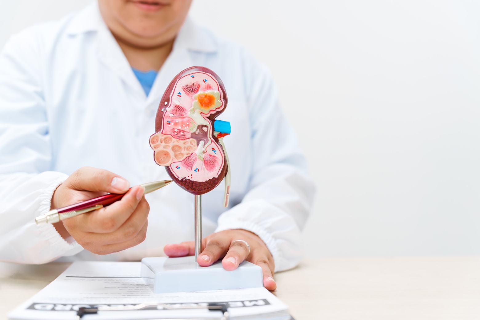
Kidney stones, also known as kidney stone disease or renal lithiasis, are a common condition characterized by the formation of solid deposits in the kidneys.
Kidney stones form when mineral salts and other substances in the urine crystallize and form ‘stones’ in the kidneys. These stones can vary in size, from small grains of sand to larger stones that can obstruct the flow of urine.
Kidney stones can cause severe pain and complications if left untreated.
Symptoms of kidney stones
The symptoms of kidney stones vary depending on the size, location, and composition of the stone. Kidney stones can be asymptomatic in its early stages, but as the stones grow or shift, they can cause severe pain and complications. The most common symptoms include:
Early symptoms:
in the initial stages, kidney stones may not present obvious symptoms. However, some patients may experience vague discomfort in the back or side.
Late symptoms:
as stones grow or move through the urinary tract, they can cause:
Intense pain in the back, side, or lower part of the abdomen (renal colic). This pain may be intermittent and can vary in intensity.
Pain that radiates to the groin or genitals.
Nausea and vomiting, which can be caused by intense pain.
Blood in the urine (hematuria), visible or microscopic.
Frequent need to urinate, even in small quantities.
Burning or pain when urinating (dysuria).
Fever and chills, which indicate a possible associated urinary tract infection.
If you experience any of these symptoms, seek medical attention for proper diagnosis and treatment.
Classification of kidney stones
Kidney stones are classified according to their chemical composition, which influences the treatment and prevention of future stones.
Calcium stones: they are the most common, representing approximately 80% of all kidney stones. They are formed mainly from calcium oxalate, but can also be composed of calcium phosphate.
Uric acid stones: they form in people with high levels of uric acid in the urine. They are more common in people with gout, a diet high in purines, or certain metabolic disorders.
Struvite stones: also known as infection stones, they form in the presence of bacteria that produce urease, an enzyme that alkalinizes the urine. They are more common in women with recurrent urinary tract infections.
Cystine stones: they are less common and form in people with cystinuria, an inherited disorder that causes excessive excretion of cystine in the urine.
Causes of kidney stones
Kidney stone formation is a complex process influenced by various factors. An imbalance in the concentration of substances in the urine triggers the formation and growth of crystals. Understanding these causes can be useful for prevention.
Certain factors increase the likelihood of developing kidney stones. Some of them can be controlled with lifestyle changes, while others cannot. These include:
Family history.
Diet: a diet high in animal protein, sodium, and oxalate may increase the risk.
Low fluid intake: drinking too little water concentrates substances in the urine, promoting the formation of kidney stones.
Obesity: overweight and obesity are associated with an increased risk of kidney stones due to metabolic alterations.
Some medical conditions: diseases such as inflammatory bowel disease, gout, hyperparathyroidism, and renal tubular acidosis increase the risk of kidney stones.
Some medications: certain medications, such as diuretics, calcium-containing antacids, and some antibiotics, can increase the risk of kidney stones.
Complications of kidney stones
If not treated properly, kidney stones can trigger various complications that affect the urinary system and overall health. These complications can vary in severity, from mild discomfort to problems requiring urgent medical intervention. The following are some of the most common complications:
Recurrent urinary infections: kidney stones can obstruct the normal flow of urine, creating an environment conducive to bacterial growth. This obstruction can make it difficult to eliminate bacteria, increasing the risk of recurrent urinary tract infections. These infections can manifest with symptoms such as burning during urination, frequent urination, fever, and pain in the lower back or side.
Urinary tract obstruction: kidney stones, especially larger ones, can block the flow of urine at any point in the urinary tract, from the kidneys to the urethra. This obstruction can cause severe pain (renal colic), inflammation, and even kidney damage if not treated quickly. The obstruction can be partial or complete, and its severity depends on the size and location of the stone. If the obstruction is complete, it may require urgent medical intervention to restore urine flow.
Kidney damage: prolonged obstruction of the urinary tract can put pressure on the kidney and damage its tissue. This damage can be reversible if the obstruction is treated in time; however, if the obstruction persists, it can cause permanent kidney damage and even kidney failure. Kidney damage can be silent in its early stages and manifest with symptoms such as fatigue, leg swelling, loss of appetite, and changes in the amount of urine.
Kidney failure: in severe cases, when urinary tract obstruction is severe or prolonged, or when there is significant kidney damage, the kidneys may lose their ability to filter waste and excess fluid from the body. This can lead to kidney failure, a serious condition that may require dialysis or a kidney transplant. Kidney failure can manifest with symptoms such as nausea, vomiting, loss of appetite, extreme fatigue, difficulty breathing, swelling, and changes in urination.
Diagnosis of kidney stones
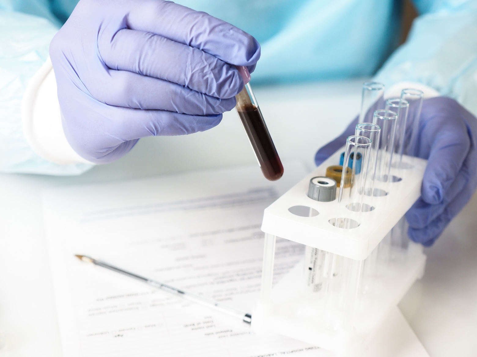
The diagnosis of kidney stones is made through different tests and examinations. These tests make it possible to determine the presence, size, and composition of the stones. The most common tests are:
Medical history and physical examination: the doctor evaluates the patient’s symptoms and general condition.
Urinalysis: it is a fundamental test for evaluating the health of the urinary tract. It can detect the presence of blood (hematuria), infection, crystals, and other abnormalities that suggest the presence of kidney stones. It also allows us to evaluate the pH of the urine, an important factor in the formation of certain types of stones.
Blood tests: blood tests complement urine tests and provide information about kidney function and the levels of certain substances that may contribute to the formation of kidney stones. We evaluate levels of calcium, uric acid, creatinine, phosphorus, and other substances to identify potential metabolic imbalances.
Imaging studies: diagnostic imaging is essential for kidney stones. Computed tomography (CT) is the most accurate diagnostic test, providing detailed images of the kidneys, ureters, and bladder, visualizing stones of any size and composition, even those not visible on X-ray or ultrasound, and detecting other abnormalities. Plain X-rays and ultrasound are also used, which can detect some stones, but with lower sensitivity. Renal ultrasound is a non-invasive test that uses sound waves to create images of the kidneys and urinary tract and detect stones of different sizes and compositions, as well as assess hydronephrosis. In some cases, an intravenous urography (IVU or Intravenous Pyelography – IVP), a contrast X-ray that shows the urinary tract and helps identify blockages caused by stones, can be performed.
Treatment of kidney stones
The treatment for kidney stones is tailored to each patient, depending on the size and location of the stone and the symptoms. The goal is to eliminate the stone or facilitate its expulsion, and to prevent the formation of new stones. Treatment options include:
Conservative measures: for small stones (less than 5 mm) that do not cause obstruction or significant symptoms, the following measures may be recommended: increased fluid intake (2–3 liters a day) to dilute the urine and facilitate the spontaneous expulsion of the stones; the use of analgesics such as paracetamol or non-steroidal anti-inflammatory drugs (NSAIDs) to relieve pain associated with the passage of stones. In cases of severe pain, opioid analgesics may be used. In turn, the use of other drugs such as alpha-blockers allows the muscles of the ureter to relax and facilitates the expulsion of the stones.
Depending on the composition of the stones, dietary changes may be recommended to reduce the risk of recurrence. For example, a diet low in sodium and animal protein may be beneficial for calcium stones, while a diet low in purines may be helpful for uric acid stones.
Surgery: for larger, symptomatic stones or those that cause complications, surgical intervention is usually the treatment of choice. There are different surgeries, and each type has its own indications, benefits, and risks, which should be discussed with the medical team.
Extracorporeal shock wave lithotripsy (ESWL): it is a non-invasive procedure that uses shock waves to fragment kidney stones into smaller pieces that can be expelled through urine. It is effective for stones up to 2 cm in diameter located in the kidney or the upper part of the ureter.
Percutaneous nephrolithotomy (PCNL): it is a minimally invasive procedure performed under general anesthesia. A nephroscope, a small instrument with a camera and light, is inserted through a small incision in the back into the kidney. Through the nephroscope, the stones can be fragmented and extracted. PCNL is effective for large or complex stones located in the kidney.
Ureteroscopy (URS): it is a minimally invasive procedure performed under general or regional anesthesia. A ureteroscope, a small tube with a camera and light, is inserted through the urethra and bladder into the ureter. Through the ureteroscope, the stones can be visualized, fragmented, and extracted. URS is effective for stones located in the ureter.
Open surgery: it is currently an uncommon procedure for the removal of kidney stones. It is reserved for complex cases or when other treatments have not been successful.
At HM Hospitales, the kidney stone unit offers a comprehensive and multidisciplinary approach to the treatment of kidney stones. Treatment options include extracorporeal shock wave lithotripsy (ESWL), percutaneous nephrolithotomy (PCNL), ureteroscopy (URS), and surgery if necessary. Our goal is to provide the most appropriate and effective treatment for each patient, minimizing the risk of complications and improving their quality of life.
Remember that this article is for informational purposes only and does not replace professional medical advice. If you suspect you have kidney stones, consult a specialist to obtain an accurate diagnosis and an appropriate treatment plan tailored to your needs.
Our doctors
Contact and make an appointment with the professionals in this area