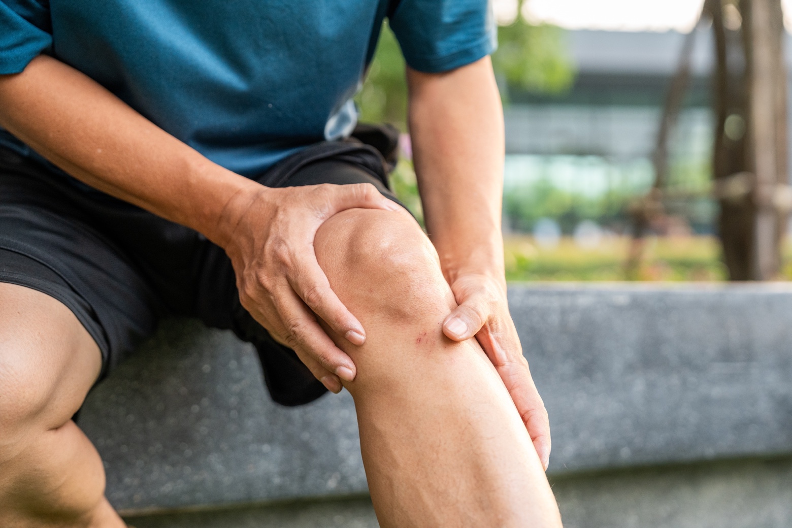
What is anterior cruciate ligament tear?
The anterior cruciate ligament (ACL) is a fundamental structure for knee stability. It connects the femur (thigh bone) to the tibia (shin bone), limiting the anterior movement of the tibia and controlling the rotation of the knee. An ACL tear occurs when this ligament tears, either partially or completely. This tear compromises the stability of the joint, increasing the risk of other injuries and making it difficult to perform activities that involve twisting or sudden changes of direction.
An ACL tear is a relatively common injury, especially in athletes and in contact sports (football, basketball, etc.). This injury can significantly affect knee stability and the ability to perform physical activities, so it is essential to understand its causes, symptoms, and treatment options.
Early symptoms:
Immediately after the injury, it is common to experience:
- Knee twisting with a “pop” or “snap” sound at the time of the injury.
- Intense and immediate pain.
- Rapid swelling of the knee.
- A feeling of instability or that the knee “is giving way.”
- Difficulty walking or bearing weight.
Late symptoms:
if the injury is not treated properly, the following symptoms may appear:
- Sensation of “locking” or “catching” in the knee.
Classification of anterior cruciate ligament tear
To determine the severity of the injury and the most appropriate treatment, ACL tears are classified into three grades according to the extent of the tear:
- Grade I: mild sprain. The ligament is stretched, but not torn. Knee stability is largely maintained.
- Grade II: moderate sprain. The ligament is partially torn, which compromises the stability of the knee.
- Grade III: complete ligament tear. The knee loses stability and is more prone to other injuries.
Causes of anterior cruciate ligament tear
Anterior cruciate ligament (ACL) tear is usually caused by sudden movements or twisting of the knee that exceed the ligament’s resistance. These situations may include:
- Changing direction quickly.
- Landing incorrectly from a jump.
- Receiving a direct blow to the knee.
Risk factors for anterior cruciate ligament tear
There are certain factors that can increase the likelihood of an ACL tear occurring. Knowing these factors can help take preventive measures.
- Sports: practicing sports that involve turns, jumps, and changes of direction, such as football, basketball, skiing, etc., significantly increases the risk.
- Sex: women have a higher risk of ACL tear than men, possibly due to anatomical and hormonal differences.
- Physical conditioning: poor physical condition or weak muscles around the knee can increase the risk.
- Inappropriate footwear: the use of footwear that does not provide sufficient support or stability.
- Playing surface: playing on uneven or slippery surfaces.
Complications of anterior cruciate ligament tear
An ACL tear that is not properly treated can lead to various complications that affect health, quality of life, and increase the risk of osteoarthritis in the joint. Approximately 3 out of 4 anterior cruciate ligament tears require surgery
- Chronic knee instability: difficulty performing activities that require stability, such as running or turning.
- Meniscus injuries: ACL tears can increase the risk of meniscus tears, structures that act as shock absorbers in the knee.
- Osteoarthritis: chronic knee instability can contribute to the development of osteoarthritis, a degenerative disease that affects articular cartilage.
- Functional limitation: difficulty performing everyday activities, such as climbing stairs or walking long distances.
Diagnosis of anterior cruciate ligament tear
Accurately diagnosing an anterior cruciate ligament (ACL) tear is essential to determining the most effective treatment plan. The diagnostic process involves a comprehensive evaluation that combines information obtained through different methods.
- Medical history: a detailed medical history is the first step in the diagnosis. The doctor will ask you about how the injury occurred, when you felt the pain, the intensity of the pain, and whether you heard any sounds (such as a pop or crack) at the time of the injury. The doctor will also ask you about your current symptoms, such as knee instability, difficulty walking, or the presence of swelling. It is important to be precise when describing your symptoms and any activities that aggravate or relieve them. This information will help the doctor understand the nature of your injury.
- Physical examination: it is essential for assessing the stability and range of motion of the knee. The doctor will carefully examine your knee, palpating the area to detect points of pain, swelling, or deformities. They will perform specific tests, such as the Lachman test, the anterior drawer test, and the pivot shift test, to assess the integrity of the ACL. These tests involve manipulating the knee in different positions to check for ligament laxity. The doctor will also assess the range of motion of your knee, both in flexion and extension, and compare the mobility of the injured knee with that of the healthy knee.
- Imaging tests: imaging tests are essential to confirm the diagnosis and rule out other possible injuries. The main imaging tests used for ACL tears are X-rays and magnetic resonance imaging (MRI). Although X-rays do not directly visualize ligaments, they are useful for ruling out bone fractures that may accompany an ACL tear. In some cases, X-rays may show an indirect sign of ACL tear, such as Segond’s sign, a small avulsion fracture on the outside of the tibia. In addition, MRI is the imaging test of choice for diagnosing an ACL tear. It provides detailed images of the soft tissues of the knee, including the ACL, menisci, and cartilage. MRI allows visualization of the extent of the ACL tear, as well as the presence of other associated injuries, such as meniscus tears or articular cartilage injuries. In some cases, MR arthrography can be used, in which a contrast medium is injected into the knee joint before the MRI to improve the visualization of internal structures.
Treatment of anterior cruciate ligament tear
ACL tear treatment is tailored to the individual needs of each patient. The decision regarding the best therapeutic approach is based on factors such as the severity of the injury, the patient’s activity level, age, the presence of other associated injuries, sports demands, and the patient’s preferences.
- Conservative treatment: it is an option for partial ruptures or for complete ruptures in people with a low level of sports activity or for those who do not wish to undergo surgery. The goal of conservative treatment is to relieve symptoms, restore knee function, and prevent future injuries.
This approach initially includes the RICE protocol (Rest, Ice, Compression, Elevation), which is applied in the acute phase of the injury to control swelling and pain. Rest involves avoiding activities that aggravate the injury. It is recommended to apply ice to the knee for 15–20 minutes every 2–3 hours. Compression with an elastic bandage helps control swelling. Keeping the knee elevated above the level of the heart also helps reduce swelling. Physical therapy is an essential component of conservative treatment. A physical therapist will guide you through specific exercises to strengthen the muscles surrounding the knee, improve stability, regain range of motion, and improve proprioception (the ability to sense the position of the knee in space). At HM Hospitales, we offer personalized physiotherapy programs for knee rehabilitation.
- Medications: painkillers and anti-inflammatories can be used to control pain and inflammation. In some cases, corticosteroids can be injected into the knee to reduce inflammation. A knee brace can be used to provide support and stability to the knee.
- Surgery: it is the treatment of choice for complete ACL tears (grade III), for active people or athletes and for those who do not respond to conservative treatment. The goal of surgery is to reconstruct the ACL since the vast majority of these injuries cannot be repaired (sutured) and to restore knee stability. ACL reconstruction surgery is usually performed by arthroscopy, a minimally invasive technique that uses small incisions and a camera to visualize and repair the injury. During surgery, the torn ligament is removed and replaced with a tissue graft. The graft can come from the patient themselves (autograft), such as the hamstring tendons or the patellar tendon, or from a donor (allograft). At HM Hospitales, we have expert surgeons in ACL reconstruction surgery, using minimally invasive techniques such as arthroscopy.
- Rehabilitation: after surgery, it is essential to follow a rehabilitation program to regain knee function. Rehabilitation includes exercises to restore range of motion, strengthen the muscles surrounding the knee, and improve proprioception.
A multidisciplinary approach, involving traumatologists, physiotherapists, rehabilitation specialists, radiologists, and other specialists, is essential for comprehensive and individualized care.
Remember that this article is for informational purposes only and does not replace professional medical advice. If you suspect you have an anterior cruciate ligament tear, consult a specialist to obtain an accurate diagnosis and an appropriate treatment plan tailored to your needs.