Brain Magnetic Resonance Imaging (fMRI)
What is functional Brain Magnetic Resonance Imaging (fMRI)?
Functional Brain Magnetic Resonance Imaging (fMRI) is an advanced neuroimaging technique that allows mapping active areas of the brain while the patient performs specific tasks. Unlike a conventional MRI that shows the brain’s structure, fMRI shows brain activity in real-time.
What is this diagnostic test used for?
fMRI is a valuable tool in:
- Neurosurgery planning: Helps identify and preserve essential brain areas (such as those for language or movement) during surgery.
- Neurological studies: Allows investigating how the brain functions in different neurological conditions, such as Alzheimer’s, Parkinson’s, or after a stroke (CVA).
- Cognitive research: Used to better understand cognitive processes such as memory, attention, and language.
Benefits of high technology in fMRI
fMRI uses a technique called BOLD (Blood Oxygen Level Dependent) imaging, which detects changes in blood oxygenation in the brain. When a brain region is active, it needs more oxygen, leading to an increase in blood flow to that area. fMRI detects these changes and visually represents them on a brain activity map.
How is the procedure performed?
The fMRI procedure involves:
-
Preparation:
You do not need special preparation for this test. You can eat and drink normally, unless your doctor tells you otherwise.
-
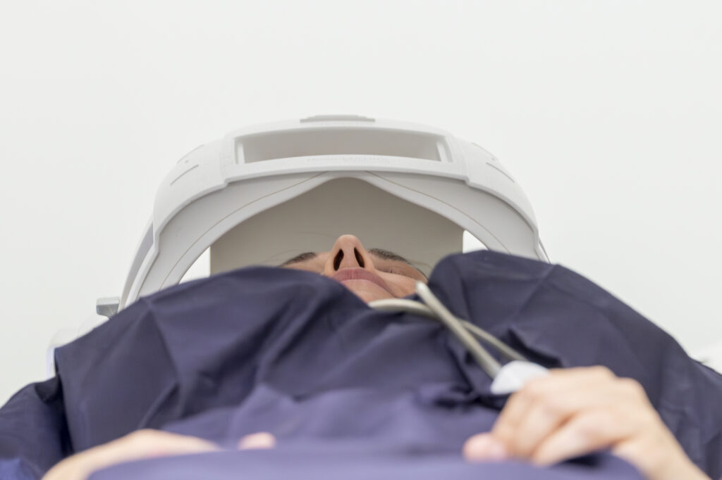
During the test:
You will be asked to lie comfortably on the MRI table. Then, special coils will be placed around your head to ensure clear images are obtained. During the test, you will be asked to perform some tasks, such as moving your hands or fingers, speaking or listening to words, or solving simple problems. It is very important that you follow the technician’s instructions and remain as still as possible while performing these tasks to obtain the best results. The duration of the test varies, but generally lasts between 45 and 90 minutes.
-
After the test:
You can resume your normal activities immediately after the test. The fMRI results will be analyzed by a radiologist and a neurologist, who will send them to your doctor. He or she will explain the findings and, if necessary, recommend appropriate treatment.
Recommendations for the test
- Remain still: It is important to remain still during the study and avoid movements to obtain clear and precise images.
- Communicate claustrophobia: If you suffer from claustrophobia, inform the staff before the test. They will offer options to help you feel more comfortable.
- Inform about metallic implants: If you have any metallic implants, such as a pacemaker, surgical clips, prostheses, or hearing aids, inform both your doctor and the MRI technician before performing the test.
Are there any risks?
fMRI is a safe and non-invasive test that does not use ionizing radiation. It does not present significant risks, but precautions are necessary in patients with pacemakers or other metallic implants. Some people may feel anxious or claustrophobic during the test. If this is your case, let us know in advance so we can help you feel more comfortable.
For your test to proceed smoothly, we ask that you arrive in advance of your scheduled time. This will allow us to complete the necessary administrative and clinical preparation.
Before the test, we will provide you with the Informed Consent form, a document with important information that you must read and sign.
If your appointment is for a Magnetic Resonance Imaging (MRI), it is crucial that you inform us about the presence of pacemakers, metallic objects, prostheses (including dental), tattoos, or medication infusion devices, such as insulin pumps.
These diagnostic tests are very safe, but as with any medical procedure, there is a minimal possibility of incidence.
