
Discover How Asthma is Treated in Adults
Asthma is a prevalent condition, affecting 2.5 million people in Spain, around 10% of the adult population. It can appea…
Pulmonary nodules are small masses of tissue in the lung, usually less than 3 centimeters in diameter, that are found on a chest X-ray or computed tomography (CT) scan. They are surrounded by healthy lung parenchyma and are not associated with atelectasis (lung collapse), enlarged lymph nodes at the hilum (the area where blood vessels and bronchi enter and exit the lung), or pleural effusion (fluid accumulation around the lung). They can be benign (noncancerous) or malignant (cancerous). If the lesion is larger than 3 cm, it is considered a lung mass, which has a higher probability of being malignant.
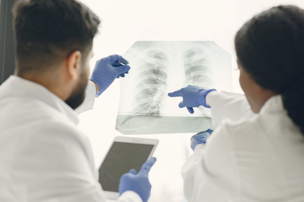
In the vast majority of cases, pulmonary nodules do not cause any noticeable symptoms. They are usually discovered incidentally during a chest X-ray or computed tomography (CT) scan of the chest performed for other health reasons. However, when the nodules are large or malignant (cancerous), some symptoms may appear, although they are often nonspecific and can be mistaken for other respiratory conditions. The most common symptoms include:
a persistent cough or one that worsens over time may be a sign of a pulmonary nodule. It can be a dry cough or a productive cough (with mucus). In some cases, especially if the nodule is irritating an airway, there may be coughing up blood (hemoptysis), although this is more common in cancerous nodules.
chest pain associated with a pulmonary nodule can be dull or sharp, constant or intermittent. It may worsen when breathing deeply or coughing. The location of the pain does not always correspond to the location of the nodule.
if a nodule is large or is blocking a main airway, it can make breathing difficult. Shortness of breath can be mild or severe, and may worsen with physical exertion.
wheezing is a whistling sound when breathing, which may indicate that the airways are inflamed or narrowed by a nodule.
in some cases, a pulmonary nodule can increase susceptibility to infections such as pneumonia or bronchitis.
It is important to note that these symptoms are not exclusive to pulmonary nodules and may be present in other respiratory diseases. If you experience any of these symptoms, especially if you smoke or have other risk factors for lung cancer, it is essential to consult a doctor for an appropriate evaluation.
Pulmonary nodules are primarily classified according to their appearance on radiological images, especially based on their density:
The classification of pulmonary nodules helps doctors assess the risk of malignancy. Subsolid nodules, especially partially solid ones, are often associated with a higher risk of lung cancer than solid nodules. Other characteristics, such as size, shape, and the presence of calcifications, are also considered in the evaluation of the nodule.
A pulmonary nodule can originate from a wide variety of causes, which often makes it difficult to characterize. Infections are a common cause, and can be bacterial, such as pneumonia or a lung abscess; fungal, such as histoplasmosis or coccidioidomycosis; or even parasitic.
Inflammatory diseases can also manifest as pulmonary nodules, with sarcoidosis and granulomatosis being common examples. Neoplasms, both benign and malignant, represent another possible cause. Benign nodules can be hamartomas or adenomas, while malignant nodules can correspond to primary lung cancer or metastases from other cancers.
In addition, there are other less frequent causes, such as arteriovenous malformations, cysts, scars from previous infections, and pulmonary embolisms. Given the diversity of possible causes, it is essential to perform a complete medical evaluation to determine the origin of the pulmonary nodule.

While most pulmonary nodules are benign, some factors increase the likelihood that a pulmonary nodule is malignant:
La The diagnostic approach to the finding of a pulmonary nodule is based on a systematic approach that combines different methods to determine its nature (benign or malignant) and its underlying cause. The diagnostic process generally includes the following steps:
The differential diagnosis of a pulmonary nodule is crucial to distinguish between the various possible causes, including infections, inflammation, benign neoplasms, and cancer. The doctor will consider all findings from the medical history, physical examination, and imaging tests to arrive at an accurate diagnosis and determine the most appropriate treatment.
The management plan for a pulmonary nodule should be individualized based on the degree of suspicion, which will depend on several factors (size, location, and characteristics of the nodule), as well as the patient’s risk factors and overall health status. Management options include:
The treatment of a pulmonary nodule should be individualized and planned by a multidisciplinary team of specialists, including pulmonologists, radiologists, thoracic surgeons, and oncologists, to ensure the best possible outcome for each patient.
Remember that this article is for informational purposes only and does not replace professional medical advice. If you suspect you have a pulmonary nodule, consult a specialist to obtain an accurate diagnosis and an appropriate treatment plan tailored to your needs.