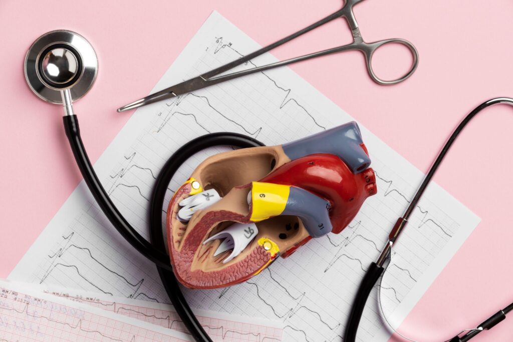
Mitral valve prolapse is a heart condition that affects the mitral valve, one of the four valves of the heart. The mitral valve is located between the left atrium and the left ventricle of the heart. Its function allows oxygen-rich blood to flow from the left atrium to the left ventricle, preventing it from flowing back.
In mitral valve prolapse, the leaflets of the mitral valve do not close properly, but bulge or prolapse into the left atrium during left ventricular contraction. This prolapse can cause some of the oxygenated blood to flow back into the left atrium, a phenomenon known as mitral regurgitation. If the regurgitation is mild, it generally does not cause problems. However, if it is severe, it can overload the heart and, over time, weaken it.
Symptoms of mitral valve prolapse
Many people with mitral valve prolapse have no symptoms, and the condition is discovered during a routine physical exam or heart test performed for another reason. When symptoms occur, they can vary widely in severity, from mild to severe. Some of the most common symptoms include:
Palpitations:
sensation of fast, strong, irregular, or “fluttering” heartbeats. They can be intermittent or last for several minutes. Sometimes, people describe the sensation as if the heart “skipped a beat,” “turned over,” or beat too fast.
Dizziness or lightheadedness:
feeling of instability or fainting.
Shortness of breath (dyspnea):
feeling of shortness of breath, especially during exertion or when lying down.
Fatigue:
feeling of tiredness or exhaustion, even after adequate rest.
Severe symptoms of mitral valve prolapse are more common in men over 50 years of age. If you experience any of these symptoms, especially palpitations, dizziness, or difficulty breathing, it is important to consult a doctor for an evaluation.
Classification of mitral valve prolapse
Mitral valve prolapse is classified into different subtypes according to characteristics such as leaflet thickness and the way the leaflets are attached to the mitral annulus:
Classic or non-classic: if the valve leaflets exceed 5 mm in thickness, it is considered classic mitral valve prolapse; Otherwise, it is non-classic.
Symmetric or asymmetric: classic mitral valve prolapse is divided into symmetric (tips of the valve leaflets joined at the same point of the annulus) and asymmetric (one leaflet is displaced further into the left atrium than the other).
Flail or non-flail: asymmetric mitral valve prolapse may be flail, with the leaflet bending back into the left atrium, increasing the risk of chordae tendineae rupture and mitral regurgitation, or non-flail. Mitral regurgitation is more common in flail asymmetric mitral valve prolapse.
Causes and risk factors of mitral valve prolapse
In most cases, the exact cause of mitral valve prolapse is unknown. It is believed that there may be a genetic predisposition, as it often runs in families. Additionally, some factors have been identified that can increase the risk of developing mitral valve prolapse, including:
Family history of mitral valve prolapse: having a close relative affected increases the risk of developing the condition.
Connective tissue diseases: certain connective tissue diseases (such as Marfan syndrome, Ehlers-Danlos syndrome, and osteogenesis imperfecta) are associated with an increased risk of mitral valve prolapse. These diseases affect the structure and strength of connective tissue, which is essential for the proper functioning of the heart valves.
Some heart diseases: diseases such as certain cardiomyopathies or coronary artery disease can increase the risk of secondary mitral valve prolapse (in this case, the prolapse is a consequence of other diseases).
In some cases, mitral valve prolapse can cause complications, although most people do not experience them. Some of the possible complications include:
Severe mitral regurgitation: if mitral regurgitation is significant, it can lead to heart failure, a condition in which the heart cannot pump enough blood to meet the body’s needs.
Infective endocarditis: it is an infection of the inner lining of the heart, usually caused by bacteria. People with disease in any heart valve have a slightly higher risk of developing it.
Cardiac arrhythmias: they are irregular heartbeats. People with mitral valve prolapse may experience different types of arrhythmias, such as atrial fibrillation, supraventricular tachycardia, or ventricular extrasystoles.
Diagnosis of mitral valve prolapse
The diagnosis of mitral valve prolapse is based on the patient’s medical history, a physical examination, and diagnostic tests. The doctor will ask about your symptoms, medical history, and family history of heart disease. During the physical exam, the doctor will listen to your heart with a stethoscope to detect a heart murmur, which may be a sign of mitral regurgitation.
The main test for diagnosing mitral valve prolapse is the echocardiogram. This test uses sound waves to create images of the heart in motion. There are different types of echocardiograms:
Transthoracic echocardiogram – Doppler (TTE): it is the most common type of echocardiogram. It is performed by placing a transducer (a microphone-like device) on the chest. The transducer emits sound waves that bounce off the structures of the heart and create images on a monitor. It also allows measurement of the speed and direction of blood flow through the mitral valve and assessment of the severity of regurgitation.
Transesophageal echocardiogram (TEE): it provides more detailed images of the heart. It is performed by inserting a probe through the esophagus, which is located behind the heart. It is used when the TTE is inconclusive or a more precise evaluation is required. It is a minimally invasive procedure that requires mild sedation.
Three-dimensional (3D) echocardiography: it reconstructs three-dimensional images of the heart and mitral valve, facilitating the understanding of valve anatomy and function. It is especially useful in surgical planning.
Cardiac catheterization: although not routinely used in the diagnosis of mitral valve prolapse, it may be necessary in some cases to assess pressure in the heart chambers and rule out coronary artery disease.
Treatment of mitral valve prolapse
The treatment of mitral valve prolapse depends on the severity of the condition and the presence or absence of symptoms. Many people with mitral valve prolapse do not require any specific treatment. The doctor may recommend regular follow-up with echocardiograms to monitor the condition.
If symptoms are present or if mitral regurgitation is severe, treatment may include:
Regular follow-up: for asymptomatic patients with mild mitral regurgitation. Follow-up includes periodic echocardiograms to monitor the progression of mitral valve prolapse and cardiac function.
Lifestyle changes: avoiding caffeine, alcohol, and tobacco, as they can worsen palpitations. Exercising regularly as directed by your doctor. Following a heart-healthy diet.
Medications: various medications can be used to control symptoms or treat complications associated with mitral valve prolapse. Beta-blockers are used to control palpitations, anxiety, and chest pain. Antiarrhythmics are used to treat cardiac arrhythmias, such as atrial fibrillation. In some specific cases and less often than in the past, prophylactic antibiotics may be prescribed before dental or surgical procedures to prevent infective endocarditis, especially in high-risk patients.
Surgery: surgery is considered in patients with severe symptomatic mitral regurgitation, severe asymptomatic mitral regurgitation with evidence of impaired left ventricular function, or complications such as infective endocarditis or rupture of the chordae tendineae. Surgical options focus on repairing or replacing the mitral valve. Mitral valve repair is the procedure of choice whenever possible, seeking to preserve the native valve and correct the prolapse using techniques such as resection of redundant valve tissue, annuloplasty (valve ring repair) or the placement of artificial chordae. This option offers better long-term results than replacement. Mitral valve replacement is performed when repair is not feasible due to the severity of the valve damage, using a mechanical prosthesis (requires lifelong anticoagulation) or a biological prosthesis (limited durability, but without long-term anticoagulation). The decision regarding the type of surgery is based on the anatomy of the valve, the surgeon’s experience, and the individual characteristics of the patient.
Remember that this article is for informational purposes only and does not replace professional medical advice. If you suspect you may have mitral valve prolapse, consult a specialist to obtain an accurate diagnosis and a treatment plan tailored to your needs.
Our doctors
Contact and make an appointment with the professionals in this area