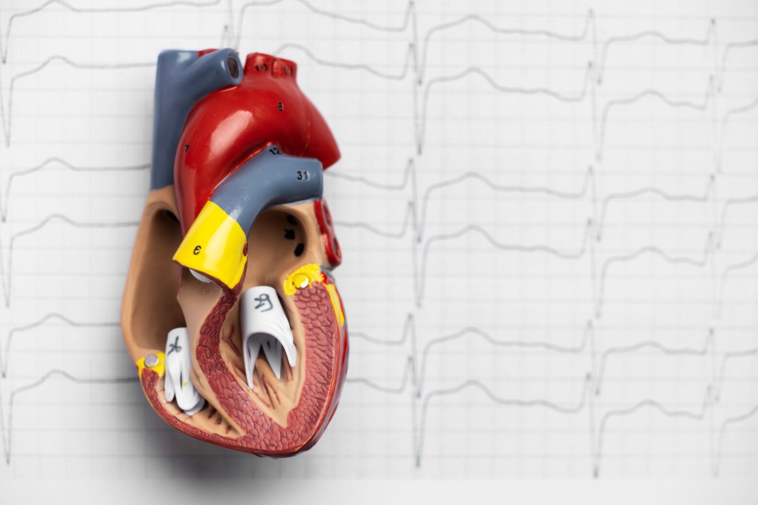
The heart has four valves that ensure blood flows in the correct direction. These four valves are the aortic, mitral, pulmonary, and tricuspid valves.
The aortic valve, located between the left ventricle (the heart’s main pumping chamber) and the aorta (the body’s largest artery), normally has three leaflets, or “flaps,” that open and close with each heartbeat. In people with a bicuspid aortic valve, this valve has only two leaflets. This malformation can affect the valve’s ability to open and close properly, which can lead to two main problems:
Aortic stenosis: the valve does not open properly, which makes it difficult for blood to flow from the heart to the aorta. This forces the heart to work harder.
Aortic insufficiency: the valve does not close properly, allowing blood to flow back into the left ventricle. This also puts extra strain on the heart.
Although most patients who experience this condition can lead a normal, symptom-free life, it is crucial to understand its potential implications and the importance of proper diagnosis and follow-up. sus posibles implicaciones y la importancia de un diagnóstico y seguimiento adecuados.
Symptoms of bicuspid valve
Many people with bicuspid valve do not experience symptoms, especially during childhood and adolescence. However, over time, the valve can deteriorate, leading to various complications and symptoms. Some of the most common symptoms include:
Shortness of breath (dyspnea):
especially during physical exercise.
Chest pain (angina):
a feeling of tightness in the chest, often triggered by physical exertion.
Fainting or dizziness (syncope):
caused by reduced blood flow to the brain.
Palpitations:
sensation of fast, strong, or irregular heartbeats.
Fatigue:
excessive tiredness and lack of energy.
It is important to note that the severity and presentation of symptoms can vary significantly from person to person. Some people may not experience any symptoms throughout their lives, while others may develop severe symptoms that require medical intervention.
Classification of bicuspid valve
The classification of bicuspid aortic valve is essential to understanding the valve anatomy, guiding treatment decisions, and predicting potential complications. There are several classification systems, but the most widely used is the Sievers classification, which is based on the number and location of the commissures and raphes (fusion lines between the valve leaflets).
Sievers classification:
Type 0: it has two symmetric leaflets with no raphe. It is the least common form of bicuspid valve.
Type 1: it has two asymmetrical leaflets with a raphe. The raphe may be located between the leaflets corresponding to the right and left commissures (R-L), the right and noncoronary commissures (R-NC), or the left and noncoronary commissures (L-NC). It is the most common type of bicuspid valve.
Type 2: it has two leaflets with two raphes. It is a less common form of bicuspid valve.
There are other classifications, such as the International Consensus, that provide a more detailed description of the anatomy of the valve and the aorta. más detallada de la anatomía de la válvula y la aorta.
Causes of bicuspid valve
Bicuspid valve is a congenital condition, meaning it is present from birth. It is believed to be due to abnormal heart development during the fetal period, around the eighth week of pregnancy. Although the exact cause is unknown, it is believed that genetic and environmental factors are involved. Mutations have been identified in certain genes that may be related to the bicuspid valve.
The main risk factor for bicuspid valve is a family history of the disease. If a close relative has the condition, the risk of developing it is higher. Currently, no other modifiable risk factors are known.
Complications of bicuspid valve
A bicuspid valve can lead to a number of complications, some of which can be serious and life-threatening. These complications include:
Aortic stenosis: the narrowing of the aortic valve, which restricts blood flow from the heart.
Aortic insufficiency: the backflow of blood into the left ventricle due to incomplete closure of the valve.
Infective endocarditis: an aortic valve infection, which can be serious and require treatment with antibiotics and/or surgery.
Aortic dilation or aneurysm: the enlargement of the aorta, which can weaken the arterial wall and increase the risk of rupture or dissection.
Heart rhythm disorders (arrhythmias): irregular heartbeats that can range from mild to life-threatening.
Diagnosis of bicuspid valve
In young patients, the diagnosis of bicuspid valve is often incidental, that is, it is discovered during a routine physical examination, an ECG performed for another reason, or due to the presence of a heart murmur detected in childhood. In these cases, echocardiography is crucial to confirm the diagnosis and assess the severity of the condition.
Echocardiogram: the diagnosis of bicuspid valve is usually made using an echocardiogram. This is a radiation-free test that uses sound waves to create images of the moving heart, allowing the cardiologist to visualize the structure and function of the aortic valve.
Multimodality imaging studies: other imaging studies, such as cardiac magnetic resonance imaging (CMR) or cardiac computed tomography (CCT), may be necessary to better visualize the aortic valve, as well as to accurately measure the dimensions of the aorta to rule out the presence of dilatation or aneurysm.
Cardiac catheterization: it is an invasive procedure that involves inserting a catheter into an artery or vein to reach the heart. Although not routinely used in the diagnosis of bicuspid aortic valve, it may be necessary to assess pressure in the heart chambers and rule out coronary artery disease.
Treatment of bicuspid valve
The treatment of bicuspid valve depends on the severity of the disease and the presence of symptoms or complications. In some cases, immediate treatment is not required, and the patient simply needs regular follow-up with a cardiologist. In other cases, medication may be recommended to control symptoms or prevent complications. If the aortic valve is severely damaged or causes significant symptoms, surgery or an interventional procedure may be necessary.
Regular follow-up: for patients with no symptoms or mild symptoms, regular follow-up with a cardiologist is essential to monitor the progression of the disease.
Medications: in aortic valve and aortic disease, medications may be necessary to achieve adequate blood pressure control or to treat arrhythmias. In some specific cases, prophylactic antibiotics may be prescribed before dental or surgical procedures to prevent infective endocarditis, especially in high-risk patients.
Surgery: surgery may be necessary to repair or replace the damaged aortic valve. Valve repair involves reconstructing the existing valve, while valve replacement consists of replacing the damaged valve with a mechanical or biological prosthesis. In some cases, it may also be necessary to repair or replace the aorta if it is dilated or has an aneurysm.
Transcatheter interventions: transcatheter aortic valve replacement (TAVR) is a minimally invasive procedure that involves inserting a new valve through a catheter that is introduced through an artery in the groin. This procedure is an alternative to open surgery for patients with high surgical risk.
Managing a bicuspid valve requires a multidisciplinary approach, involving cardiologists, cardiac surgeons, cardiac imaging specialists, and other healthcare professionals.
Remember that this article is for informational purposes only and does not replace professional medical advice. If you suspect you have complications and symptoms related to bicuspid valve, consult a specialist to obtain an accurate diagnosis and an appropriate treatment plan tailored to your needs. nsulta con un especialista para obtener un diagnóstico preciso y un plan de tratamiento adecuado a tus necesidades.
Our doctors
Contact and make an appointment with the professionals in this area