PET-MRI Scan
What is a PET-MRI scan?
PET-MRI is an advanced hybrid imaging technology that combines Positron Emission Tomography (PET) and Magnetic Resonance Imaging (MRI). This combination allows metabolic and functional images of the body (through PET) to be obtained along with high-resolution anatomical images (through MRI), providing a more accurate and complete diagnosis in a single scan.
What is this procedure for?
PET-MRI is primarily used in:
- Oncology: it is used for the diagnosis, staging, and monitoring of malignant tumors. It is especially useful for identifying small or hard-to-detect lesions.
- Neurology: it is used to evaluate neurodegenerative diseases such as Alzheimer’s, epilepsy and other neurological conditions.
- Cardiology: it is used to analyze blood flow and cardiac function.
- Medical research: widely used in clinical studies and in the evaluation of new therapies.
Benefits of advanced technology in PET-MRI
PET uses radiopharmaceuticals that emit positrons to detect metabolic activity in the body, while MRI uses magnetic fields and radio waves to generate detailed images of internal structures. By combining both techniques, PET-MRI provides anatomical, functional, and metabolic information in a single study, improving diagnostic accuracy.
What does the procedure involve?
The PET-MRI procedure involves:
-
Preparation:
You will generally need to fast for 4 to 6 hours before the scan, but check with your doctor to confirm. It is recommended to drink water before the procedure. Be sure to tell your doctor if you are taking any medication, if you are pregnant, or if you are breastfeeding. Also, wear comfortable clothing that does not contain metal components.
-
During the procedure:
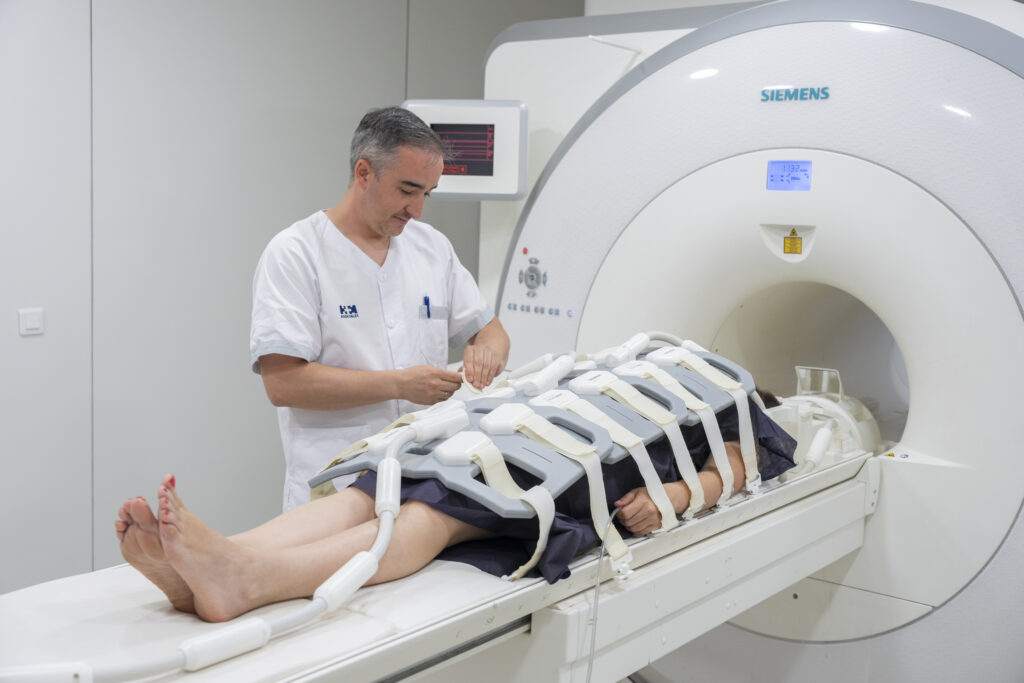
During the procedure, a radiopharmaceutical will be administered to you intravenously. This substance is safe and is quickly eliminated from the body due to its short half-life. Next, you will be asked to lie down on the PET-MRI scanner table. First, metabolic images will be taken using the PET technique, and then anatomical images will be taken using MRI. It is important that you remain still during the study to ensure the quality of the images. The procedure usually lasts between 60 and 90 minutes.
-
After the procedure:
After the procedure, you will be able to resume your normal activities, unless your doctor advises otherwise. It is advisable to drink plenty of water to help eliminate the radiopharmaceutical from your body. The results of the PET-MRI scan will be analyzed by a specialist radiologist and sent to your doctor, who will explain the findings and recommend the appropriate treatment, if necessary.
Recommendations for the procedure
During the procedure, it is important to remain as still as possible to ensure the accuracy of the images. In addition, follow all medical instructions before, during, and after the procedure to ensure optimal results.
Are there any risks?
PET-MRI is a safe procedure, but there are some minor risks:
- Exposure to radiation: PET uses a small amount of radiation due to the administered radiopharmaceutical. However, the dose is low and considered safe.
- Claustrophobia: some people may experience anxiety while inside the MRI scanner. Inform the medical team if this happens to you; steps can be taken to help you feel more comfortable.
- Allergic reactions (rare): in exceptional cases, allergic reactions to the radiopharmaceutical may occur.
To ensure your procedure runs smoothly, we ask that you arrive before the scheduled time. This will allow us to complete the necessary administrative and clinical preparation.
Before the procedure, we will give you the Informed Consent form, a document with important information that you must read and sign.
If your appointment is for an MRI, it is crucial that you inform us about the presence of pacemakers, metallic objects, prostheses (including dental prostheses), tattoos, or drug infusion devices such as insulin pumps.
These diagnostic tests are very safe, but as with any medical procedure, there is still the unlikely possibility of an incident.
Do you need to undergo this procedure?
Make an appointment