Digital Breast Tomosynthesis (DBT)
What is digital breast tomosynthesis (DBT)?
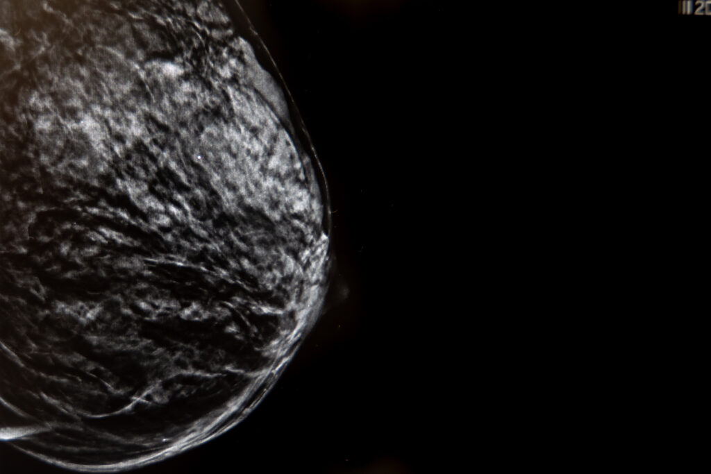
Digital breast tomosynthesis, also known as a 3D mammography, is an advanced breast imaging technique that uses low-dose X-rays to create a three-dimensional image of the breast. Unlike traditional mammography, which provides a two-dimensional image, tomosynthesis captures multiple images from different angles, allowing for a more detailed visualization of the breast tissue.
What is this diagnostic procedure for?
The main uses of digital breast tomosynthesis are in:
- Breast cancer screening: It allows the identification of small tumors that might go unnoticed in a conventional mammogram, especially in breasts with high tissue density.
- Greater precision in dense breasts: It reduces tissue overlap, improving the visualization of breast structures in women with dense breasts.
- Reduction of false positives: It reduces the need for additional testing by decreasing the number of suspicious images.
- Detailed lesion assessment: It allows for a more accurate assessment of the detected lesions, helping to determine whether they are benign or malignant.
- Early detection: It is a very useful tool in the early diagnosis of breast cancer.
Benefits of advanced technology in digital breast tomosynthesis
A digital breast tomosynthesis offers a number of key benefits thanks to the technology it uses:
- 3D images: It provides a 3D visualization of the breast, allowing for better detection of lesions.
- Low dose of radiation: It uses a dose of radiation comparable to that of a conventional digital mammogram.
- Advanced digital processing: It uses advanced software to reconstruct images and improve the visualization of breast tissue.
Detection of hidden lesions: It helps detect lesions that might not be seen in a traditional mammogram.
What does the procedure involve?
A digital breast tomosynthesis is similar to a conventional mammography, but with some differences:
-
Preparation:
You will be asked to remove the clothing on your upper body. It is important to know that you will not undergo the test if you are pregnant.
-
During the procedure:
First, you need to stand in front of the mammography machine. Next, the breast is positioned on a platform and compressed with a clear plastic paddle. In tomosynthesis, the X-ray tube moves in an arc over the breast, capturing multiple images from different angles. The process takes several seconds for each breast.
-
After the procedure:
The procedure lasts 10 to 15 minutes in total. If you have implants, it is likely that more images will be taken of each breast, making the procedure somewhat longer. The images are then analyzed by a specialized radiologist.
Recommendations for the procedure
Remember that it is important to adhere to the following recommendations to ensure the quality of the test and your comfort:
- Do not use deodorants, creams, lotions, or talc on your armpits or breasts.
- Bring previous studies (if any) so the radiologist can compare them. If you have it done at HM, you don’t need to provide them, as they will already have them on file.
- The breasts are less sensitive one week after menstruation, but the procedure is very well tolerated by most patients.
- Please advise if you are pregnant or breastfeeding, as you will likely be advised to undergo another test.
- The results will be available in less than a week or sometimes even on the same day. Ask at the end of the procedure.
Are there any risks?
Digital breast tomosynthesis is generally safe, but like any X-ray study, there are some minimal risks to consider:
- Exposure to radiation: It uses a low dose, similar to a conventional mammography. The benefit of early detection far outweighs the risk of radiation.
- Discomfort during the procedure: The compression may be uncomfortable, but it only lasts a few seconds. If you have sensitive breasts, try scheduling the test after your menstrual period.
- Possible false positives: It can detect suspicious images that may lead to additional testing or unnecessary biopsies. However, this risk is lower than with conventional mammography.
- Although it improves cancer detection, it is not able to detect all types of cancer. It is often recommended to complement the study with a breast ultrasound and/or breast MRI.
It is a safe, accurate, and recommended method for the early detection of breast cancer, and the benefits far outweigh the risks.
To ensure your procedure runs smoothly, we ask that you arrive before the scheduled time. This will allow us to complete the necessary administrative and clinical preparation.
Before the procedure, we will give you the Informed Consent form, a document with important information that you must read and sign.
If your appointment is for an MRI, it is crucial that you inform us about the presence of pacemakers, metallic objects, prostheses (including dental prostheses), tattoos, or drug infusion devices such as insulin pumps.
Do you need to undergo this procedure?
Make an appointment