Contrast-enhanced mammography
What is a contrast-enhanced mammography (CEM)?
A contrast-enhanced mammography, also called a contrast-enhanced spectral mammography (CESM), is an advanced breast imaging technique that combines digital mammography with intravenous administration of an iodinated contrast agent and advanced digital processing. Its objective is to analyze the vascularization of the breast and highlight abnormal areas or masses of tissue that may take up more contrast, a sign of tumor activity.
What is this diagnostic procedure for?
The main uses of contrast-enhanced mammography are in:
- Breast cancer screening: In breasts with a high density of glandular and fibrous tissue, cancer may be more difficult to detect on a regular mammogram. CEM increases tumor visibility by highlighting their vascularization.
- Characterization of suspicious lesions: When a mammogram or other imaging technique detects an abnormality in the breast, but the results are inconclusive, CEM can help determine whether it is a benign or malignant lesion.
- Local staging of breast cancer: Following a cancer diagnosis, CEM helps determine if there are other lesions in that breast or in the contralateral breast, allowing for a better definition of the extent of the disease.
- Monitoring response to cancer treatments: It allows the effectiveness of chemotherapy to be monitored before surgery, assessing whether the tumor is responding to treatment.
- Follow-up with patients with personal and family history of breast cancer: In high-risk patients, CEM can be a valuable tool for early detection, complementing other tests.
- Alternative to magnetic resonance imaging (MRI) of the breast: For patients who cannot undergo an MRI or find it difficult to tolerate, CEM is an alternative.
Benefits of advanced technology in CEM
A contrast-enhanced mammography offers a number of key benefits thanks to the technology it uses:
- Improved detection in dense breasts: It facilitates the early detection of cancer in breasts with high density, where conventional mammograms may be less effective.
- Greater diagnostic accuracy: It allows for a more accurate differentiation between benign and malignant lesions, reducing the need for unnecessary biopsies.
- Functional information: It provides information about the vascularization of the lesions, which helps to determine their activity and aggressiveness.
- Treatment planning: It facilitates more accurate cancer staging, helping to plan the most appropriate treatment for each patient.
Monitoring the response to treatment: It allows for the assessment of whether the tumor is responding to treatment and for more informed decisions to be made.
What does the procedure involve?
A contrast-enhanced mammography is a quick and well-tolerated test that is similar to conventional mammography, but with some important differences:
-
Preparation:
It is essential to inform your doctor if you have an allergy to iodine or radiological contrast media, suffer from any kidney disease, have thyroid problems, are pregnant, or are breastfeeding. Furthermore, before the test, you must remove all metal objects (jewelry, piercings, etc.) and avoid using deodorants, creams, or talc in the breast and armpit area. For this type of test, it is not necessary to have fasted beforehand.
-
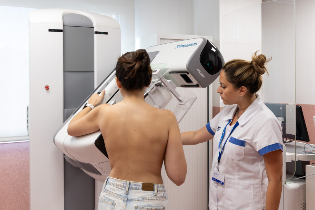
During the procedure:
A nurse will insert an IV into a vein in your arm to administer the iodinated contrast agent. You will be positioned in front of the mammography machine, and your breast will be briefly compressed between two plates. Next, several images will be taken of each breast, first without contrast and then with contrast. The complete scan usually only takes about 10 minutes. It is important to note that a feeling of warmth or a strange sensation when the contrast is injected is normal, so do not worry.
-
After the procedure:
Drink plenty of water to help eliminate the contrast from your body. You can resume your normal activities immediately after the procedure. However, if you experience itching, rash, swelling, or difficulty breathing, see a doctor to rule out a possible allergic reaction.
Recommendations for the procedure
Remember that it is important to adhere to the following recommendations to ensure the quality of the test and your comfort:
- Report allergies: it is crucial to inform the medical staff if you have a known allergy to iodine or any contrast medium.
- Inform about pregnancy or breastfeeding: If you are pregnant or breastfeeding, you should inform your doctor before the test, as the contrast medium may not be suitable in these situations.
- Report kidney problems: If you have known kidney problems, it is important to inform your doctor, because the contrast medium is eliminated through the kidneys.
- Avoid using deodorants, talcum powder, or lotions: Do not use these products on the chest or underarm area on the day of the test, as they may interfere with the images.
- Inform your doctor of any medication you are taking: This is especially necessary if you take diabetes medication (such as metformin), as it may require adjustments before or after the test.
.
Are there any risks?
A contrast-enhanced mammography is generally considered safe, but like any medical procedure involving the administration of a contrast medium, there are some potential risks:
- Allergic reactions to the contrast medium: Although uncommon, allergic reactions can occur, ranging from mild (itching, rash) to severe (difficulty breathing, anaphylactic shock). The medical staff is prepared to handle these situations.
- Contrast-induced nephropathy (CIN): In patients with compromised kidney function, the contrast medium can damage the kidneys. Special precautions will be taken if you have a history of kidney problems.
- Exposure to radiation: Although the dose of radiation in a mammogram is low, there is a minimal risk associated with radiation exposure. The benefits of early detection of breast cancer generally outweigh this risk.
- Discomfort or pain at the injection site: Some people may experience pain, bruising, or swelling at the site where the contrast medium is injected.
To ensure your procedure runs smoothly, we ask that you arrive before the scheduled time. This will allow us to complete the necessary administrative and clinical preparation.
Before the procedure, we will give you the Informed Consent form, a document with important information that you must read and sign.
If your appointment is for an MRI, it is crucial that you inform us about the presence of pacemakers, metallic objects, prostheses (including dental prostheses), tattoos, or drug infusion devices such as insulin pumps.
Do you need to undergo this procedure?
Make an appointment