Artro RM
What is MRA?
Magnetic resonance arthrography (MRA) is an advanced imaging test that allows for a highly detailed study of the joints. It is a very useful technique for diagnosing problems that are not easily identified with a conventional MRI scan.
MRA is a magnetic resonance imaging scan in which a small amount of contrast agent is introduced directly into the joint. This contrast helps the internal structures of the joint, such as the ligaments, cartilage, and labrum, to be seen much more clearly.
What is this diagnostic procedure for?
MRA is used to:
- Diagnose ligament injuries, such as tears or sprains.
- Detect problems in the articular cartilage, such as wear or injuries.
- Evaluate the labrum, a structure that helps stabilize some joints, such as the shoulder and hip.
- Identify problems in the tendons surrounding the joint.
- Assess the condition of the joint after surgery.
- Detect loose bodies within the joint.
Benefits of the latest technology in MRA
MRA combines magnetic resonance imaging technology with intra-articular contrast injection:
- High-field magnetic resonance imaging: It uses a powerful magnet to obtain high-resolution images of the joint.
- Specialized imaging sequences: It uses imaging sequences designed to visualize the internal structures of the joint in great detail.
Intra-articular contrast: The contrast agent injected into the joint helps to make the structures much clearer, which facilitates the diagnosis of injuries.
The MRA procedure is split into two parts:
-
Contrast injection:
First, you will be injected with a contrast medium into the joint to improve the quality of the images. To do this, the skin of the area to be studied will be carefully cleaned and a local anesthetic will be applied to reduce any discomfort. Next, a thin needle will be inserted into the joint and the contrast agent will be administered. In some cases, radiology or ultrasound equipment will be used to ensure the needle is in the correct position.
-
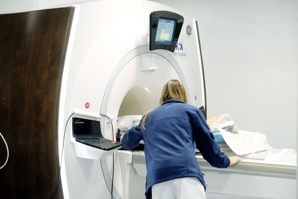
Magnetic resonance imaging:
Once the injection has been performed, the magnetic resonance imaging will be performed. You will need to lie down on the MRI table and remain as still as possible so clear images can be obtained. During the procedure, the equipment will emit some sounds, but the medical staff will provide you with hearing protection if necessary. The test can last between 30 and 60 minutes, depending on the joint being evaluated and the number of images needed for diagnosis.
Recommendations for the procedure
To ensure the study is conducted safely and effectively, please keep the following in mind:
- You need to inform your doctor if you have an allergy to the contrast agent or any medication.
- You need to inform your doctor if you are pregnant or think you might be.
- If you are taking anticoagulants (blood thinners), consult your doctor to see if you need to stop taking them before the test.
- On the day of the test, you should wear comfortable clothing and avoid wearing jewelry or other metal objects.
Are there any risks?
MRA is a safe test, but like any medical procedure, it carries some risks:
- Pain or discomfort: You may feel pain or discomfort in the joint after the injection. These symptoms are usually mild and go away within a few days.
- Infection: There is a small risk of infection in the joint after the injection.
- Allergic reaction to the contrast agent: This is uncommon, but it can happen.
- Bleeding: In rare cases, minor bleeding may occur in the joint after the injection.
To ensure your procedure runs smoothly, we ask that you arrive before the scheduled time. This will allow us to complete the necessary administrative and clinical preparation.
Before the procedure, we will give you the Informed Consent form, a document with important information that you must read and sign.
If your appointment is for an MRI, it is crucial that you inform us about the presence of pacemakers, metallic objects, prostheses (including dental prostheses), tattoos, or drug infusion devices such as insulin pumps.
