3D Carotid and Femoral Vascular Ultrasound
What is a three-dimensional carotid and femoral ultrasound?
A three-dimensional carotid and femoral ultrasound is an ultrasound technique that uses 3D imaging to evaluate the carotid arteries (located in the neck) and the femoral arteries (located in the legs). This procedure can detect abnormalities such as atherosclerotic plaques, narrowing (stenosis), or changes in blood flow, which are key for preventing serious cardiovascular diseases such as stroke or peripheral artery disease.
What is this procedure for?
A three-dimensional carotid and femoral ultrasound is mainly used for:
- Atherosclerosis detection: to detect fatty or calcified plaque buildup in the arteries.
- Cardiovascular risk assessment: it is used to determine the risk of a stroke or heart attack.
- Diagnosis of arterial stenosis: it helps detect narrowing of the arteries that may impede blood flow.
- Clinical follow-up: it is used to monitor the progression of vascular diseases and evaluate the effectiveness of treatments.
- Treatment planning: it helps plan procedures such as angioplasty or vascular surgery.
Benefits of advanced technology in a three-dimensional carotid and femoral ultrasound
The procedure uses a transducer that emits ultrasonic waves to generate three-dimensional images of the carotid and femoral arteries. These images allow for analysis of arterial wall structure, identification of atherosclerotic plaques, and real-time measurement of blood flow.
The carotid and femoral three-dimensional ultrasound procedure involves:
-
Preparation:
To prepare, the doctor will review your medical history and cardiovascular risk factors. It is recommended that you wear comfortable clothing, as you will need to expose your neck and legs to facilitate the examination. Also, if you are taking anticoagulants or other cardiovascular medications, inform the medical team so they can take this into account.
-
During the procedure:
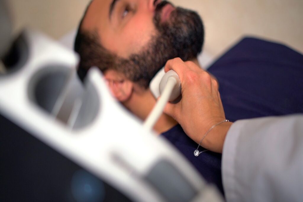
During the procedure, a gel will be applied to the areas to be examined (neck and legs) to facilitate the transmission of the ultrasonic waves. The transducer will be gently moved over the carotid and femoral arteries to obtain detailed three-dimensional images. In some cases, color Doppler will be used to assess the speed and direction of blood flow. The procedure usually lasts between 30 and 60 minutes.
-
After the procedure:
You can resume your normal activities immediately after the procedure. A cardiologist specializing in this field will analyze and interpret the results and send them to your doctor.
Recommendations for the procedure
It is important that you remain as still as possible during the procedure to ensure the quality of the images. Be sure to follow all medical instructions before, during, and after the procedure to ensure accurate results.
Are there any risks?
The three-dimensional carotid and femoral vascular ultrasound is a safe, non-invasive procedure. It does not involve exposure to radiation or require medication to be administered. However, there may be some minor considerations:
- Feeling of cold: from the conductive gel applied to the skin.
- Mild discomfort: if pressure is applied with the transducer in sensitive areas.
To ensure your procedure runs smoothly, we ask that you arrive before the scheduled time. This will allow us to complete the necessary administrative and clinical preparation.
Before the procedure, we will give you the Informed Consent form, a document with important information that you must read and sign.
If your appointment is for an MRI, it is crucial that you inform us about the presence of pacemakers, metallic objects, prostheses (including dental prostheses), tattoos, or drug infusion devices such as insulin pumps.