3D Echocardiography
What is a 3D Echo Cardio?
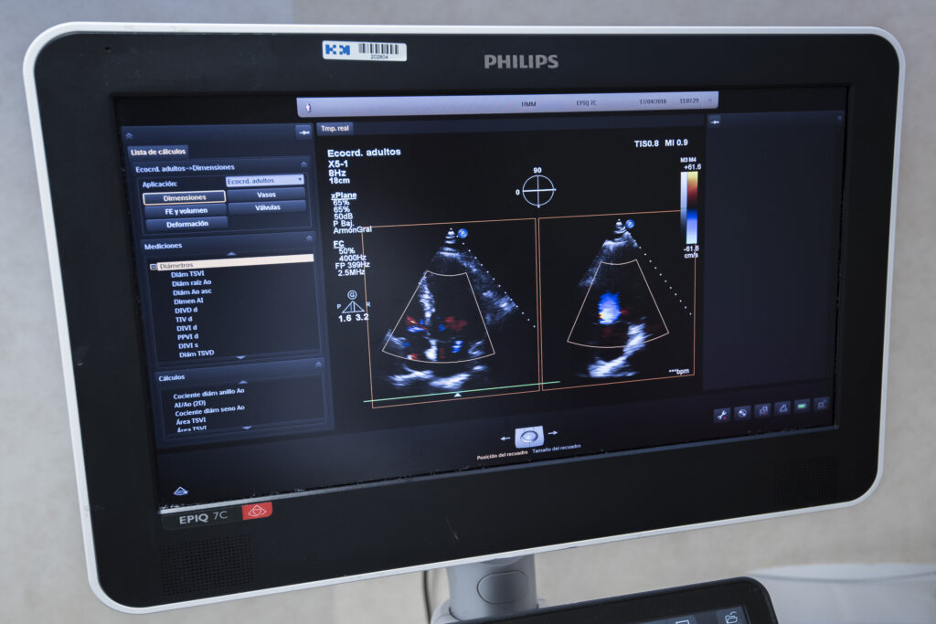
Three-dimensional echocardiography (3D Echo Cardio) is an advanced cardiac imaging technique that uses ultrasound to obtain three-dimensional images of the heart. Unlike conventional two-dimensional echocardiography, this technology allows for a more detailed and precise visualization of cardiac structures, providing more complete information about their anatomy and function.
What is this test for?
3D Echo Cardio is primarily used for:
- Evaluation of valvular diseases: Serves to analyze alterations in valves such as the mitral, aortic, or tricuspid, including stenosis and insufficiency.
- Diagnosis of congenital heart defects: Helps identify structural malformations in pediatric and adult patients.
- Surgical planning: Facilitates surgeons in planning procedures such as valve repairs or replacements through detailed visualization.
- Intraoperative monitoring: Provides real-time images during cardiac surgical interventions.
- Evaluation of ventricular dysfunction: used to study the function of the left and right ventricle, including the analysis of volumes and ejection fraction.
- Clinical research: Serves to expand knowledge about cardiovascular diseases and evaluate new therapies.
Benefits of High Technology in 3D Echo Cardio
3D Echo Cardio uses specialized transducers that emit ultrasonic waves to capture volumetric images of the heart. These images are processed by advanced software to generate three-dimensional reconstructions that allow for the analysis of cardiac structures from different angles.
What does the procedure involve?
The 3D Echo Cardio procedure involves:
-
Preparation:
For preparation, the cardiologist will evaluate your medical history and symptoms. It is advisable to wear comfortable clothing, as you will need to expose your chest to facilitate transducer placement. Additionally, if you have a pacemaker or other implanted electronic device, inform the medical team so they can take it into account.
-
During the test:
During the test, you will lie on a stretcher in a comfortable position. A gel will be applied to your chest to improve the transmission of ultrasonic waves. Then, the doctor will move a transducer over your chest to capture three-dimensional images of your heart. In some cases, a transesophageal transducer may be used to obtain more detailed images. The images will be processed in real-time and stored for later analysis. The procedure usually lasts between 30 and 60 minutes.
-
After the test:
After the test, you can resume your normal activities immediately. A specialized cardiologist will analyze and interpret the results and send them to your doctor.
Recommendations for the test
It is important to remain as still as possible during the test to ensure image quality. Additionally, follow all medical instructions before, during, and after the procedure to ensure an accurate result.
Are there any risks?
3D Echo Cardio is a safe and non-invasive procedure. However, some specific tests may cause mild discomfort:
- Transient discomfort: If a transesophageal transducer is used, you might feel discomfort in your throat due to the insertion of the device.
- Cold sensation: Due to the conductive gel applied to the chest.
For your test to go smoothly, we ask that you arrive in advance of your scheduled time. This will allow us to complete the necessary administrative and clinical preparation.
Before the test, we will provide you with the Informed Consent form, a document with important information that you must read and sign.
If your appointment is for a Magnetic Resonance Imaging (MRI), it is crucial that you inform us about the presence of pacemakers, metallic objects, prostheses (including dental ones), tattoos, or medication infusion devices, such as insulin pumps.
